Typical magnetic resonance imaging scan showing the coracohumeral
Por um escritor misterioso
Last updated 16 novembro 2024


A 61-year-old man with adhesive capsulitis of the shoulder. (AeC) MRI
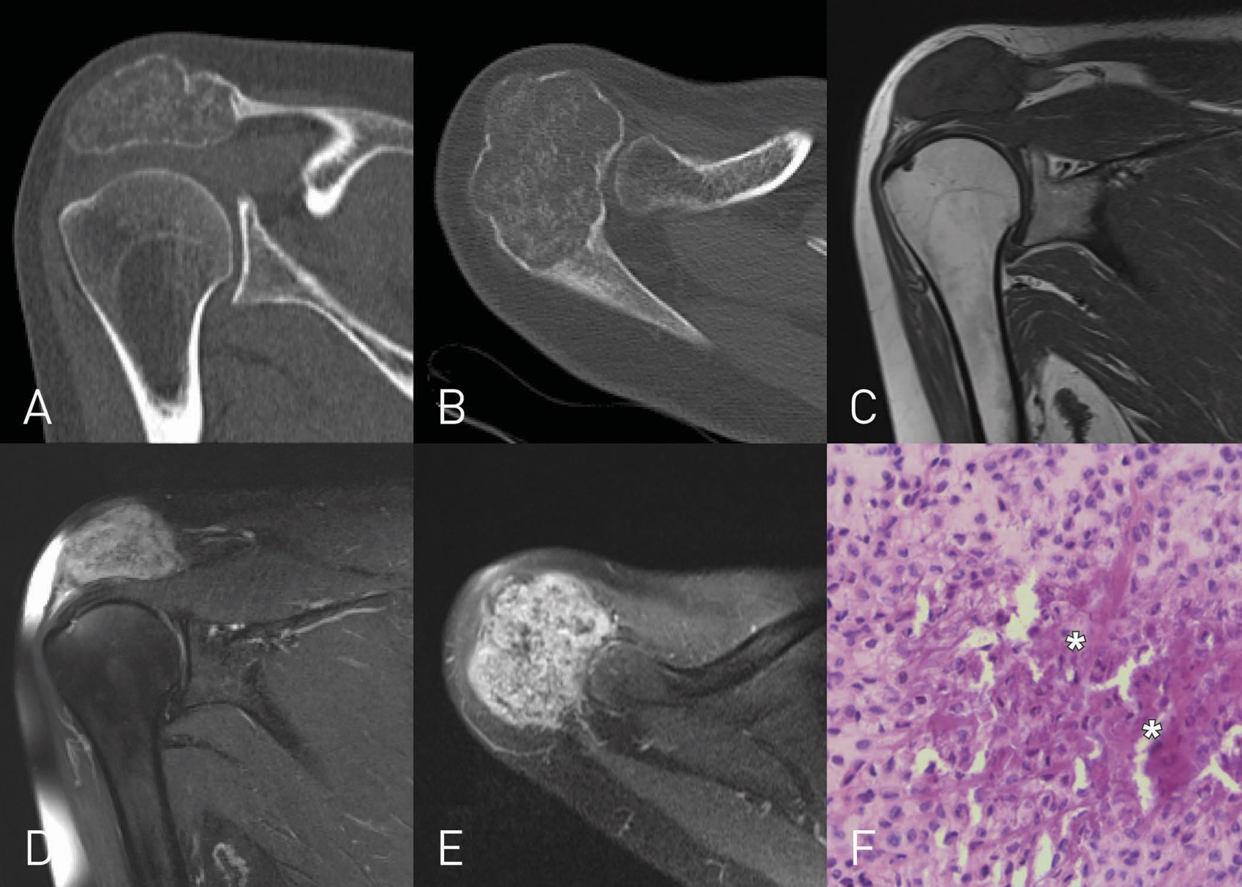
A 38-Year-Old Man with Long-Term Shoulder Pain - JBJS Image Quiz
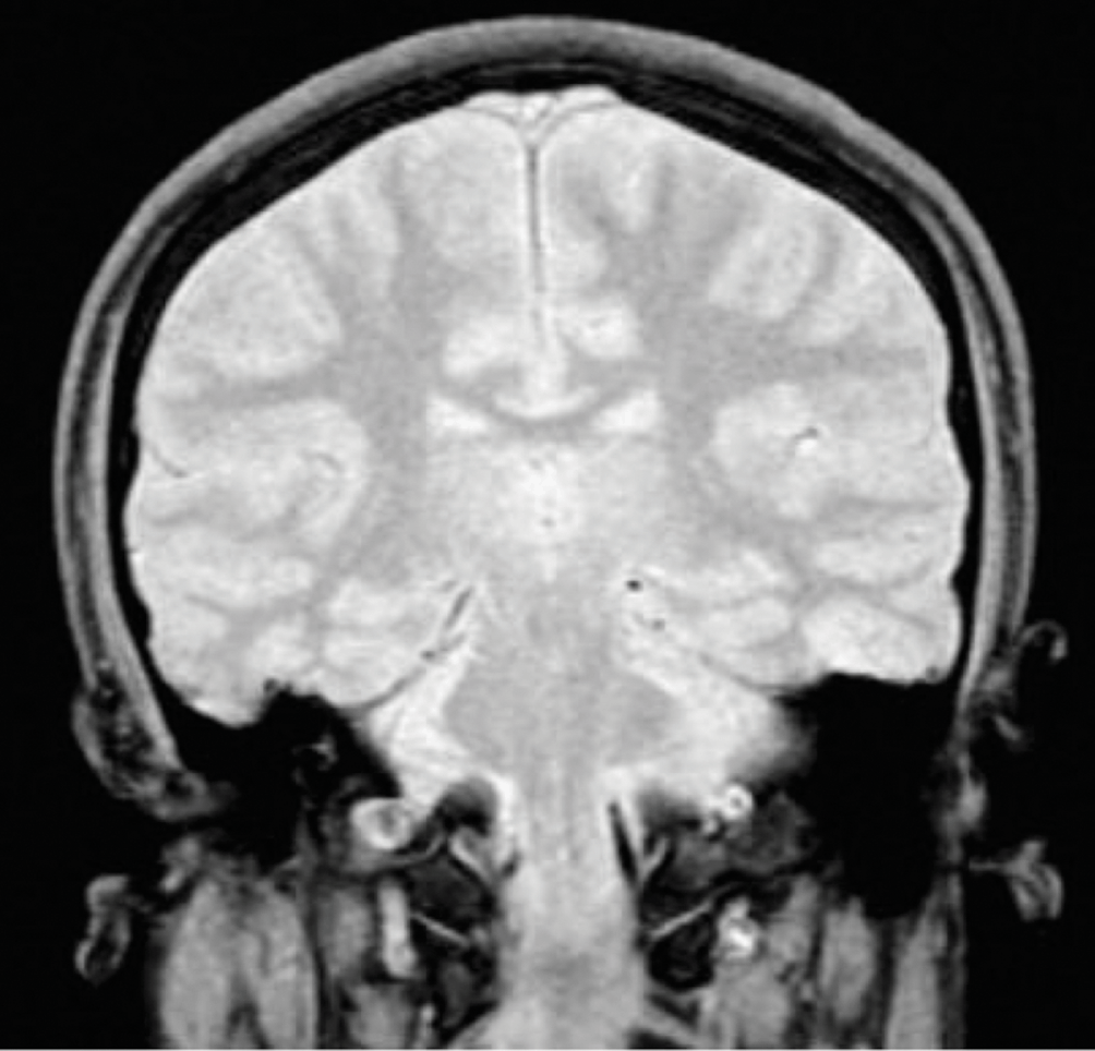
Magnetic Resonance Imaging (Part IV) - Clinical Emergency Radiology

Pain related to rotator cuff abnormalities: MRI findings without clinical significance - Bencardino - 2010 - Journal of Magnetic Resonance Imaging - Wiley Online Library

Shoulder Anatomy and Normal Variants - Journal of the Belgian Society of Radiology

Shoulder MRI: normal anatomy

Narrowed coraco-humeral distance on MRI: Association with subscapularis tendon tear - ScienceDirect
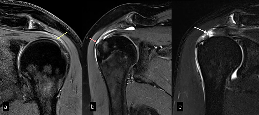
Cureus, Role of Magnetic Resonance Imaging in the Evaluation of Rotator Cuff Tears

Imaging of the Acromioclavicular Joint: Anatomy, Function, Pathologic Features, and Treatment
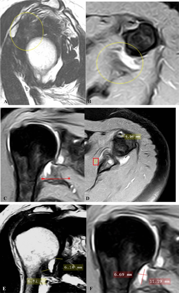
Shoulder adhesive capsulitis: can clinical data correlate with fat-suppressed T2 weighted MRI findings?, Egyptian Journal of Radiology and Nuclear Medicine
Recomendado para você
-
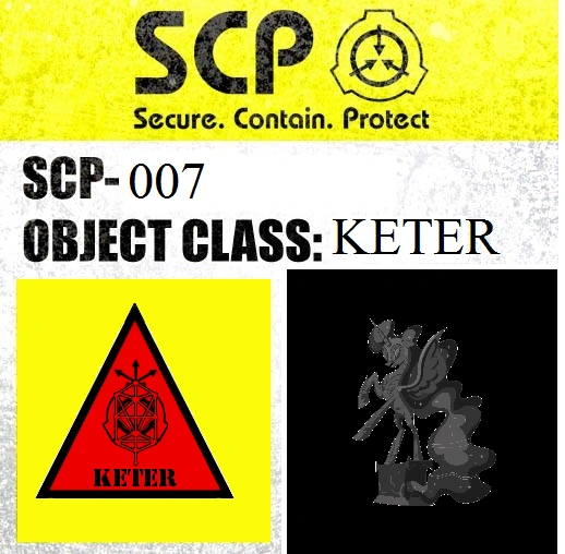 SCP-007, SCP: Containment is Magic Wiki16 novembro 2024
 -
 Abdominal Planet - SCP-00716 novembro 2024
 -
 ArtStation - SCP-007 , Ran Guo16 novembro 2024
 -
 SCP 007 : r/SCP16 novembro 2024
 -
 scp-007 fan art (this one was challenging) : r/SCP16 novembro 2024
 -
 SCP-007-J - Drawception16 novembro 2024
 -
 The SCP Foundation - Casual Cards - Yugioh Card Maker Forum16 novembro 2024
 -
 SCP-007-J - Двоечник, Вселенная SCP terra51 вики16 novembro 2024
 -
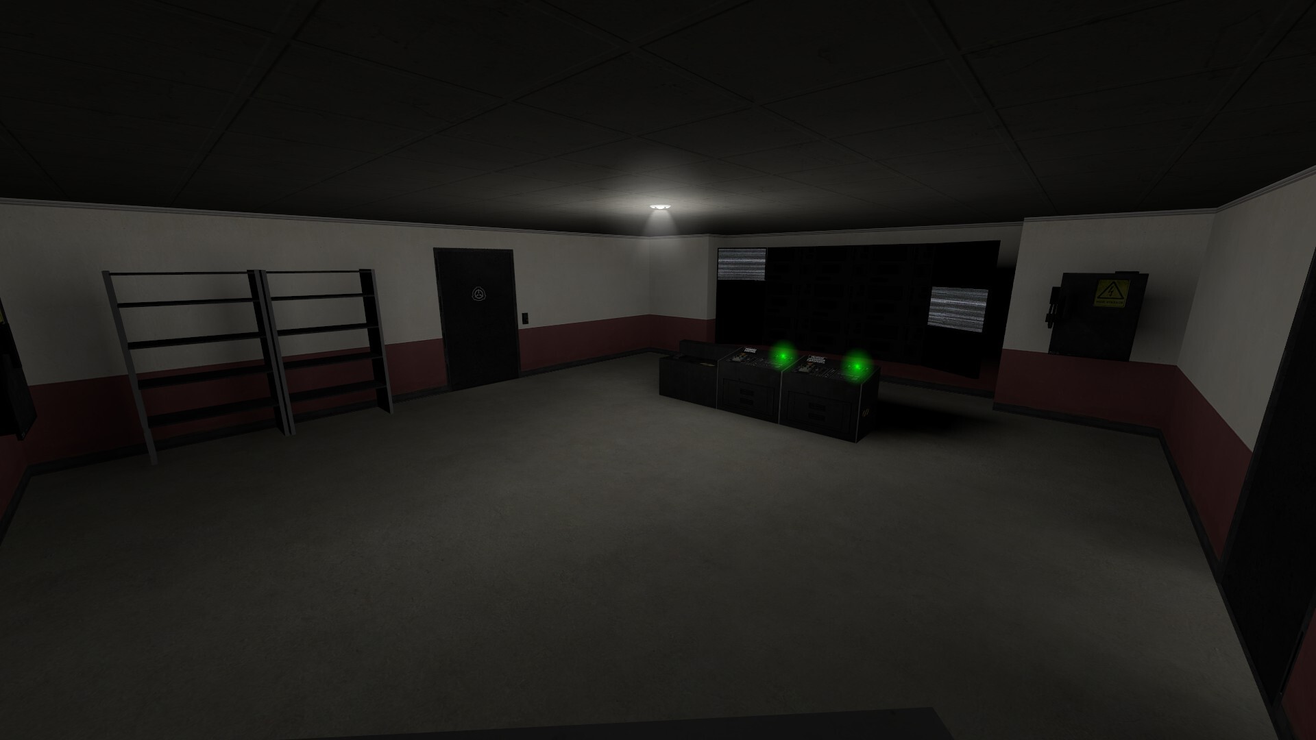 ArtStation - Source-engine Level Design16 novembro 2024
 -
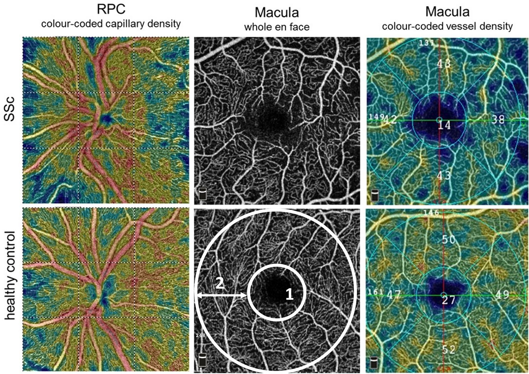 Altered ocular microvasculature in patients with systemic sclerosis and very early disease of systemic sclerosis using optical coherence tomography angiography16 novembro 2024
você pode gostar
-
 Gta Grand Theft Auto Liberty City Stories - Game PLAYSTATION Psp Complete Tbe16 novembro 2024
 -
 Coastal Jewelry Women's 'True Love Waits' Cursive Script Stainless Steel Ring16 novembro 2024
 -
 Kuroko no Basket: Oshaberi Shiyou ka (Completo) – Peak Spider Fansub16 novembro 2024
 -
DMARGE - Pierce ready to take on this week. Irish actor Pierce16 novembro 2024
-
 Five Nights at Freddy's: SL v2.0 IPA : Scott Cawthon : Free Download, Borrow, and Streaming : Internet Archive16 novembro 2024
 -
 Arifureta Shokugyou De Sekai Saikyou Season 2 - What We Know So Far16 novembro 2024
 -
 Halloween - A Noite do Terror (1978) Filmes clássicos de terror, Filmes antigos de terror, Cartazes de filmes de terror16 novembro 2024
 -
 Suporte Para Agachamento Sumô Musculação Fitness Academia16 novembro 2024
 -
 Ik the first 2 we're already over. But not a lot of people in16 novembro 2024
 -
 JOGO da VELHA para 2 em COQUINHOS16 novembro 2024
