Figure 1 from Brain surface temperature under a craniotomy.
Por um escritor misterioso
Last updated 15 novembro 2024

Fig. 1. Rapid cooling of the brain surface in an in vivo mouse preparation. A: schematic representation of a cranial window during recording of temperature and single-cell activity in the anesthetized mouse. The main potential routes of heat transfer are indicated. B: brain surface temperature measured with the thermocouple during replacement of the artificial cerebrospinal fluid (ACSF) with fresh ACSF warmed to 38°C. ACSF was replaced twice, indicated by the arrowheads. - "Brain surface temperature under a craniotomy."
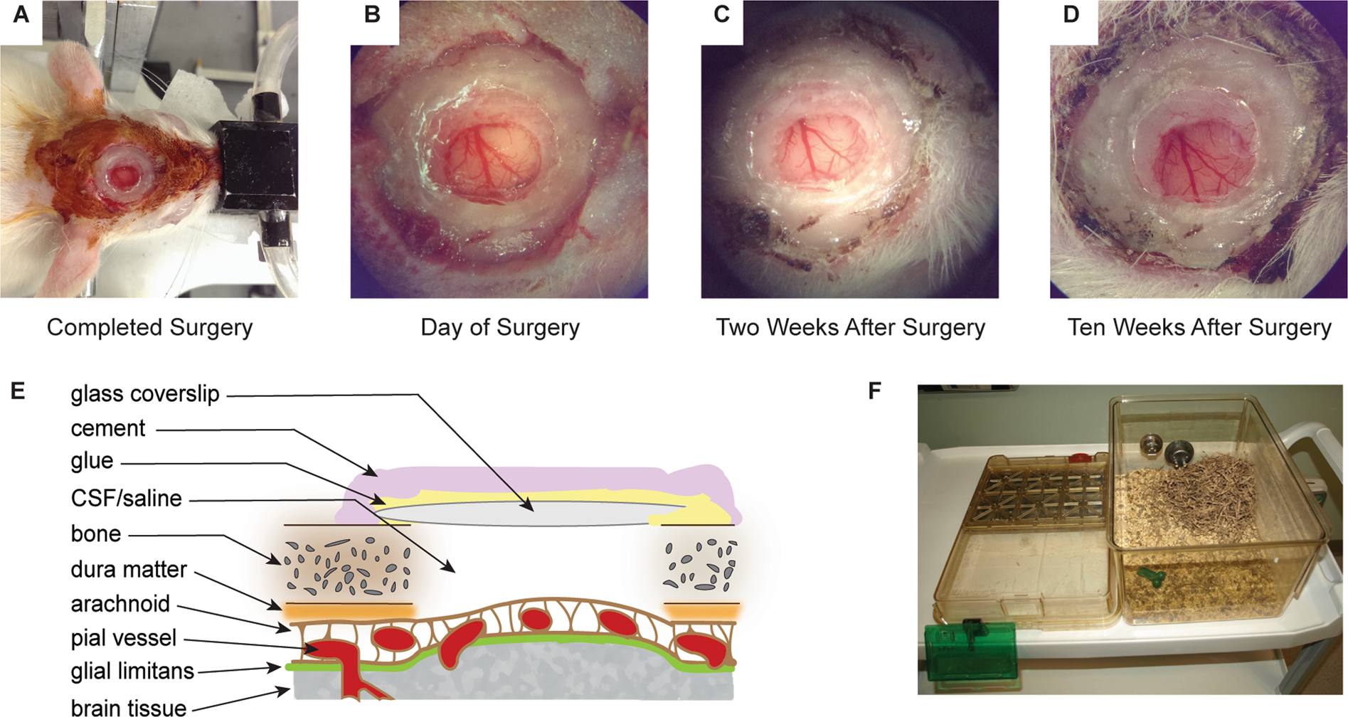
Refinement of a chronic cranial window implant in the rat for longitudinal in vivo two–photon fluorescence microscopy of neurovascular function

Data collection and craniotomy. Left: The infrared camera setup is

IJERPH, Free Full-Text

Surgical procedure for the MCA ligation. (A) After retraction of the

Figure 1 from Brain surface temperature under a craniotomy.
Quantitative third-harmonic generation imaging of mouse visual cortex areas reveals correlations between functional maps and structural substrates
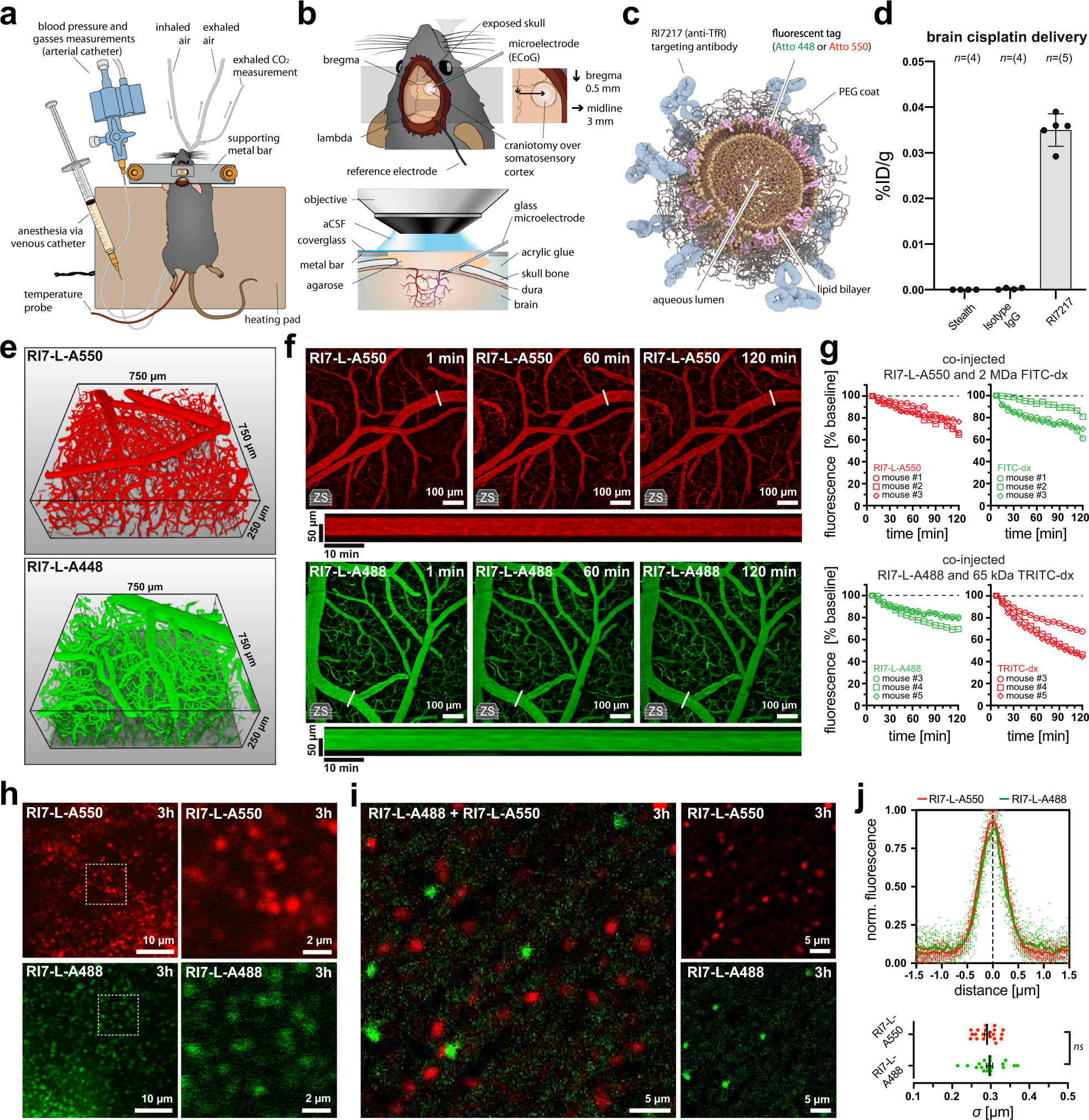
Post-capillary venules are the key locus for transcytosis-mediated brain delivery of therapeutic nanoparticles

A focal brain-cooling device as an alternative to electrical stimulation for language mapping during awake craniotomy: patient series in: Journal of Neurosurgery: Case Lessons Volume 2 Issue 2 (2021) Journals

Figure 1 from Brain surface temperature under a craniotomy.

Increase in high-gamma power with increased task difficulty in the

Brain surface temperature under a craniotomy

Cranial window for longitudinal and multimodal imaging of the whole mouse cortex
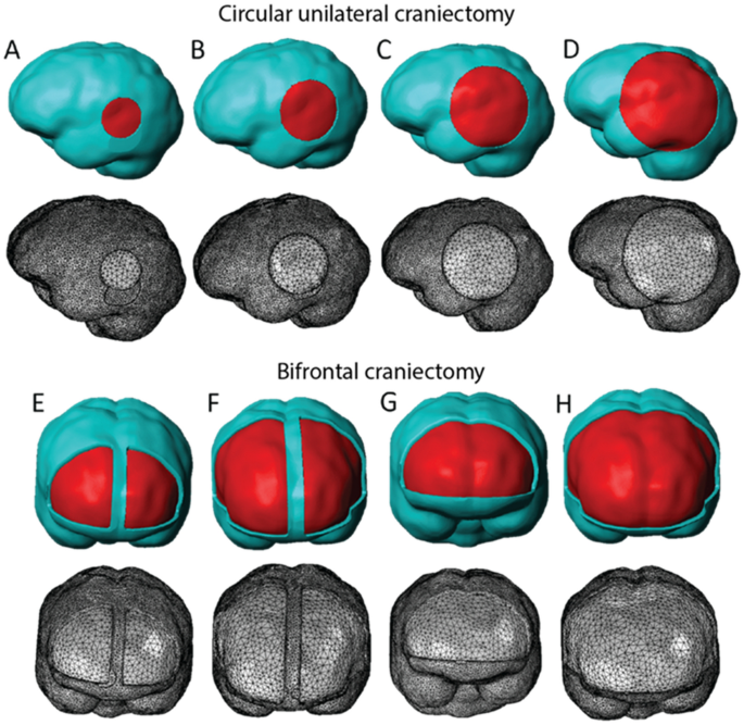
Decompressive craniectomy of post-traumatic brain injury: an in silico modelling approach for intracranial hypertension management

Full article: Brain temperature and its role in physiology and pathophysiology: Lessons from 20 years of thermorecording
Recomendado para você
-
 BRAIN TEST NÍVEL 367 EM PORTUGUÊS15 novembro 2024
 -
 Kunci Jawaban Brain Test Level 361 362 363 364 365 366 367 368 369 370: Saatnya Mencari Cuan - Tribunbengkulu.com15 novembro 2024
 -
 Brain Test Уровень 367 ответы (Он хочет большие мышцы)15 novembro 2024
 -
Solved Question 14 9 pts Listed below are brain volumes15 novembro 2024
-
Performance of Brain-Injured versus Non-Brain-Injured Individuals on Three Versions of the Category Test - Page 120 - UNT Digital Library15 novembro 2024
-
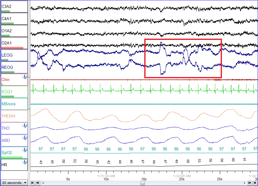 Polysomnography - Wikipedia15 novembro 2024
 -
 CapCut_sua nota instagram o4315 novembro 2024
 -
A Cell Type Selective YM155 Prodrug Targets Receptor-Interacting Protein Kinase 2 to Induce Brain Cancer Cell Death15 novembro 2024
-
Enfamil Gentlease Ready To Use Bottles - 8 Fl Oz Each/6ct : Target15 novembro 2024
-
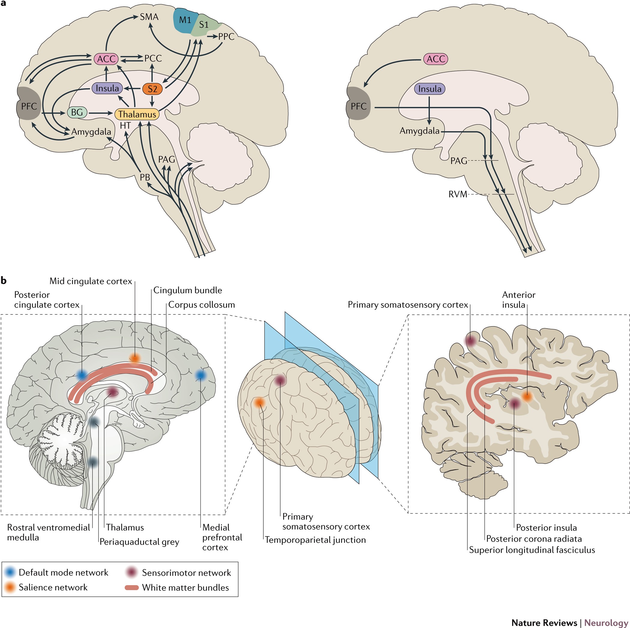 Brain imaging tests for chronic pain: medical, legal and ethical15 novembro 2024
você pode gostar
-
 Dominus Dudes, Jazwares Roblox Toys Wiki15 novembro 2024
 -
 Revista Anime Clube by Yug Jorge - Issuu15 novembro 2024
 -
 Quebra-cabeça Jogo De Quebra-cabeça Para Crianças Com Cão15 novembro 2024
 -
Jogos de Vestir Bonecas15 novembro 2024
-
 FL Studio 21 Signature Bundle Edition Complete Home studio15 novembro 2024
 -
 20 jogos grátis na Steam pra você passar o tempo na quarentena, Página: 315 novembro 2024
 -
![Stream VHS Sans - Phase 2 [Better Start Running] [Original] by](https://i1.sndcdn.com/artworks-T6ZNQm45IO5pcHki-gs1FaQ-t500x500.jpg) Stream VHS Sans - Phase 2 [Better Start Running] [Original] by15 novembro 2024
Stream VHS Sans - Phase 2 [Better Start Running] [Original] by15 novembro 2024 -
 The Last American Vampire - Wikipedia15 novembro 2024
 -
 Use of passkeys expands as passwordless authentication push advances15 novembro 2024
 -
 Download GTA San Andreas Apk + Mod v2.00 android 202115 novembro 2024


